Medications
Key Focus Areas for Pre-Oxygenation Before Moderation Sedation
#health #rescue #sedation #sedation training basics
Author:
Dulce E. Boucher
Oct 31, 2023
Back to blog
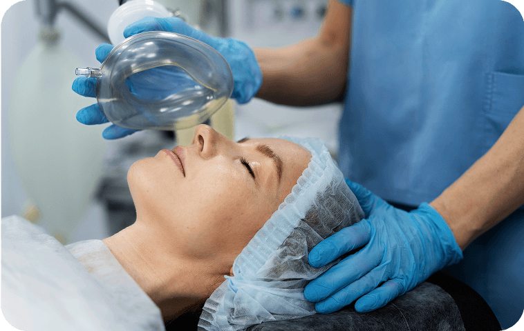
Pre-oxygenation is an important technique for guarding patients against oxygen desaturation and hypoxia during apnea with sedation. It is performed by increasing the percentage of oxygen that patients breathe during procedures. It can be accomplished by exposing patients to higher levels of oxygen via a nasal cannula, face mask, non-rebreather, or other appropriate breathing devices.
When patients receive higher levels of oxygen during procedures, rather than simply breathing room air, their reserve of oxygen in the lungs is increased. This is crucial because although the goal during moderate sedation is to maintain adequate patient respirations and responsiveness, sedation is a continuum and it is always possible for a patient to unexpectedly transition from a minimal sedation state to a deeper one, potentially losing the ability to breathe independently.
When a patient who is only breathing room air does become apneic, whether it is from airway obstruction, oversedation, or some other complication, oxygen reserves are often limited, and desaturation can occur within seconds. By pre-oxygenating with supplemental oxygen, the time to desaturation after apnea can be prolonged, giving sedation providers more time to recognize apneic episodes, prompt the patient to take a deep breath, or initiate provider rescue ventilation before oxygen desaturation occurs.
Equipment and Medications Needed for Preoxygenation
Pre-oxygenation can be accomplished using various types of equipment to provide supplemental oxygen therapy to patients during procedures. During moderate sedation, patients are exposed to medications that can result in apnea, so it is also vitally important to ensure that emergency airway equipment and reversal medication agents are available to counteract the effects of apnea.
The equipment and medications necessary to provide pre-oxygenation are described below.
Basic pre-oxygenation equipment:
Nasal cannula, simple face mask, or non-rebreather mask
Oxygen tank with regulator
Pipeline supply with flow meter
Source of suction
Suction tubing, catheters/yankauers
Oral and nasal airways
Emergency equipment:
Self-inflating bag valve mask
Advanced airway management equipment (for practitioners with intubation skills)
Laryngoscope blades and handles
Endotracheal tubes of various sizes
Pharmacologic antagonists:
Naloxone
Flumazenil
The choice of equipment and medication will always depend on the individual peculiarities of the patient and their condition.
Assessing Oxygen Level and Ensuring Proper Positioning
To ensure that pre-oxygenation will be efficient and safe for a given patient, it is advisable to evaluate their baseline oxygen saturation, assess cardiovascular vital signs, and complete medical history before beginning. Additionally, pre-oxygenation is most efficiently accomplished with optimal patient positioning and will be described in more detail below.
Baseline Oxygenation Assessment
Most healthy patients will have a baseline oxygen saturation of 95-100% on room air. This may seem like an adequate starting point, however, even for healthy patients with no comorbidities, medications used to achieve sedation are known to occasionally result in both ventilatory depression and oxygen desaturation.
In addition, some medical conditions can make patients more susceptible to low oxygen saturation levels, further predisposing them to apnea and hypoxia during sedation. An example of underlying diseases known to be risk factors include obesity, obstructive sleep apnea, heart disease, and chronic obstructive pulmonary disease.
For maximum safety, a baseline set of vital signs and a complete medical history should be performed on each patient before pre-oxygenation and sedation. With this basic information, sedation providers will be equipped to anticipate a patient’s potential need for higher levels of supplemental oxygen during procedures. Maintaining pre-oxygenation throughout sedation can help to avoid desaturation and maintain patient parameters as near to baseline as possible.
Best Positions for Efficient Pre-Oxygenation
Consideration should be given to proper positioning during procedures to improve or maintain oxygenation. If possible, patients should be placed in positions where the head is slightly elevated, rather than fully supine, as this will result in the most optimal pre-oxygenation. An even elevation of only 20 degrees can improve pre-oxygenation results.
If a patient is in the supine, lateral, head-down, or prone position during a procedure and experiences ventilatory depression or hypoxia that does not improve by other means, it is always advisable to halt the procedure, consider calling for additional help, and check that your monitors are accurate.
You must then consider moving the patient into the supine or head-up position until oxygenation and ventilation are improved and decide if reversal agents should be given and whether the procedure can be safely continued.
Supplemental Oxygen Administration
If it is anticipated that a patient may develop hypoxemia during sedation, supplemental oxygen should be administered. Oxygen administration during sedation can be delivered in various ways and functions by allowing the patient to breathe a mixture of 100% oxygen and room air. This mixture of oxygen and entrained air can be manipulated to provide a variable percentage of oxygen to the patient by adjusting the flow rate of oxygen that is delivered.
Ways to Administer and Flow Rates Management
Choosing the right equipment to deliver supplemental oxygen is based on individual patient and procedure characteristics. Things to consider include, patient comorbidities and expected need for oxygen therapy, patient comfort and tolerance for varying supplemental oxygen delivery systems, and oxygenation equipment that may interfere with the procedure.
For example, facemasks may not be able to be used in some procedures like endoscopy, due to blockade of the mouth. In that case, a low- or high-flow nasal cannula may be more appropriate. Examples of delivery systems to improve oxygenation during sedation include but are not limited to the following:
Low-flow nasal cannula
Simple face mask
Non-rebreathing mask with a reservoir can achieve oxygen levels of 90% with flow rates of 10 to 15 L/minute.
High-flow nasal cannula
Low-Flow Nasal Cannula
The traditional nasal cannula is placed in the patient’s nares and can achieve oxygen levels of approximately 35-45% by increasing flow rates up to 4 to 6 liters per minute. The nasal cannula is an open system that allows for the leaking of air into the breathing system.
To increase the percentage of oxygen delivered, the flow rate must also be increased. However, at higher flow rates the nasal mucosa can be irritated and lead to drying or bleeding so is therefore limited to these lower rates.
Simple Face Mask
A simple face mask has no bag attached and can achieve oxygen levels of approximately 35-40% with flow rates of 6 to 10 liters per minute. The mask covers the mouth and nose more fully, therefore the amount of air that leaks in around the oxygen is decreased.
This allows for higher patient exposure to oxygen. The disadvantage is that because the mouth is blocked, it cannot be used for procedures that occur through the mouth.
Non-Rebreather Mask
Non-rebreathing masks can achieve oxygen levels of approximately 90% with flow rates of 10 to 15 liters per minute. These masks have a reservoir bag attached as well as one-way valves and exhalation ports, which allow for the delivery of 100% oxygen and minimize the amount of air that leaks into the system.
The disadvantage is that because the mouth is blocked, it cannot be used for procedures that occur through the mouth.
High-Flow Nasal Cannula make this font the same as the Non-Rebreather Mask
A high-flow nasal cannula is placed in the patient’s nares and can achieve oxygen levels of 21%-100% by increasing flow rates up to 60 liters per minute. The components of the high-flow nasal cannula include an oxygen and air blender, a humidifier, and temperature control via heated tubing. These elements allow for better control of oxygen delivery and patients can tolerate much higher flow rates.
Patient Monitoring During Pre-Oxygenation
Patient monitoring during sedation is crucial for reducing adverse clinical outcomes and improving patient satisfaction. Standard monitoring recommendations include observing the patient’s level of responsiveness, pulmonary ventilation, oxygenation, and hemodynamics. This can be achieved through direct patient observation as well as the use of electrocardiogram, blood pressure, capnography, and pulse oximetry monitors.
Direct Patient Observation
Providers should continuously assess the patient for signs and symptoms of inadequate ventilation and oxygenation. This includes awareness of the patient’s level of sedation, consciousness, and comfort, their pattern and rate of respirations, and any changes in the patient’s skin color to pale or blue, which could indicate low blood pressure or hypoxia.
Throughout preoxygenation, patients should remain able to respond purposefully to verbal commands to be able to breathe when prompted and maintain adequate patterns of respiration.
Pulmonary Ventilation and Oxygenation
Clinical observation alone is often inadequate to detect apnea and hypoxemia. Therefore, monitors such as pulse oximetry and capnography should be used continuously during sedation and preoxygenation.
Of note, oxygen desaturation can be a late sign of poor ventilation. Capnography, however, is more sensitive for detecting apneic episodes and should be used simultaneously with pulse oximetry monitoring to help detect and prevent patient hypoxia.
Cardiovascular Monitoring
Non-invasive blood pressure, heart rate, and electrocardiographic monitoring are important for detecting the effects of sedatives given during sedation and preoxygenation and can facilitate early detection of adverse cardiovascular events.
Blood pressure and heart rate should be monitored continuously. The electrocardiogram is highly recommended for all patients, but not required for healthy patients without cardiovascular disease.
Poor Oxygenation: First Signs and Troubleshooting
Should a patient become apneic, there will be a variable amount of time for sedation providers to recognize it and respond before oxygen saturation levels decrease. Recognizing potential poor oxygenation is done through a combination of direct patient observation and assessing data from the monitors.
If a sedation provider is paying close attention, the potential for poor oxygenation can be recognized first by visual cues from the patient. This includes decreased or absent chest rise, snoring or paradoxical breathing, and altered level of consciousness or responsiveness. During this stage, the patient may be having inadequate respirations or apnea, but with a prompt response from the sedation provider may never show signs of poor oxygenation on pulse oximetry.
The next most likely indication of the potential for poor oxygenation will be by a change in the capnographic waveform showing lower, less frequent, or even absent waveforms. These waveforms can indicate obstruction, hypoventilation, or apnea respectively.
Even before changes in pulse oximetry are registered, the capnographic waveform can alert a sedation provider to poor ventilation which, left untreated, may eventually lead to poor oxygenation. Capnography is especially useful when patient positioning, draping, or lighting makes it difficult to adequately visually assess patients and should be used in parallel with direct observation.
Keep in mind that like any monitor, the capnographic waveform can be misleading if misplaced. For example, if a patient’s capnographic waveform is absent on the monitor, but the patient is awake and breathing without difficulty, it could be that the monitor fell off or the tubing is kinked and is not actually reading the patient’s expiratory CO2.
This type of misinterpretation can occur with any of the standard monitors. Therefore, it is imperative that sedation providers always assess the patient first and then confirm the accuracy of the monitors.
Finally, by the time poor oxygen saturation is demonstrated on pulse oximetry, oxygen levels are already at dangerously low levels and likely to decrease rapidly. Therefore, oxygen saturation monitoring should not be a first-line monitor for apnea. Instead, capnography and visual inspection of the patient will help you to more quickly diagnose a ventilatory problem.
High-Risk Patients
As part of a thorough preparation for moderate sedation, providers should be familiar with each patient’s preoperative medical history and determine if any conditions predispose the patient to low oxygen saturation levels or who may be at risk of rapid desaturation should apnea occur.
Examples of conditions that predispose patients to lower oxygen levels or rapid desaturation include:
Cyanotic heart disease
Congestive heart failure
Anemia
Obstructive lung disease (COPD, ILD, etc.)
Obesity
Pregnancy
States of elevated oxygen consumption (pediatrics, fever, severe pain, etc.)
One method that can be used for determining risk and appropriateness for moderate sedation is the ASA physical status classification system. This system uses a patient’s medical comorbidities as part of the assessment for periprocedural risk and can be used in combination with other indicators of patient risk such as procedure type, patient frailty, and conditioning.
The ASA classification is one part of the patient evaluation that can help determine if moderate sedation is appropriate, or if a higher level of care, such as monitored anesthesia care (MAC), is warranted.
Conclusion
Pre-oxygenation of patients is vital for the prevention of oxygen desaturation. It helps to mitigate the risk of hypoxemia and ensure successful moderate sedation procedures. Pre-oxygenation is simple and achieved by placing supplemental oxygen via nasal cannula, face mask, or non-rebreather on all patients throughout all procedures requiring sedation.
Especially for patients whose baseline comorbidities may predispose them to rapid desaturation, it is imperative to determine the most appropriate level of supplemental oxygen to begin with. At the same time, it’s important to recognize that higher levels of oxygen can always be achieved by escalating the method of oxygen delivery or by increasing oxygen flows.
By exposing all patients to higher levels of oxygen than what is found in room air, the time to desaturation with apnea increases and allows more leeway for providers to recognize and respond appropriately to the apneic patient.
With proper monitoring including pulse oximetry, ECG, blood pressure, and capnography, sedation providers can remain attentive to the patient and rapidly recognize when a problem arises. Remember that it is important to always assess the patient first (responsiveness, chest rise, fogging in the mask, skin color, etc.) and then assess the monitors for accuracy.
By continuously monitoring the patient for responsiveness and adequate respirations, paying close attention to the capnographic waveform, and providing supplemental pre-oxygenation during procedures, oxygen desaturation can be prevented.
About the author

Dulce E. Boucher, MD is an assistant professor at the University of Utah, board certified in Anesthesiologyand Pain Medicine. She has a particular interest in patient and provider wellness as well as medical education. In addition to her time in the operating room as an anesthesiologist, she serves part of her time as a core faculty educator for medical students at the University of Utah School of Medicine.